
RHIZARTHROSE
Arthrose des Daumensattelgelenks

WHERE IS THE SADDLE JOINT?
The saddle joint is formed by the first metacarpal bone and the trapezium bone in the wrist, whose articular surface is saddle-shaped. It is the joint that allows the thumb to be spread and opposed to the other fingers. This enables the grasping and holding of objects, both large and small.
WHAT IS THUMB SADDLE JOINT ARTHRITIS AND HOW DOES IT MANIFEST?
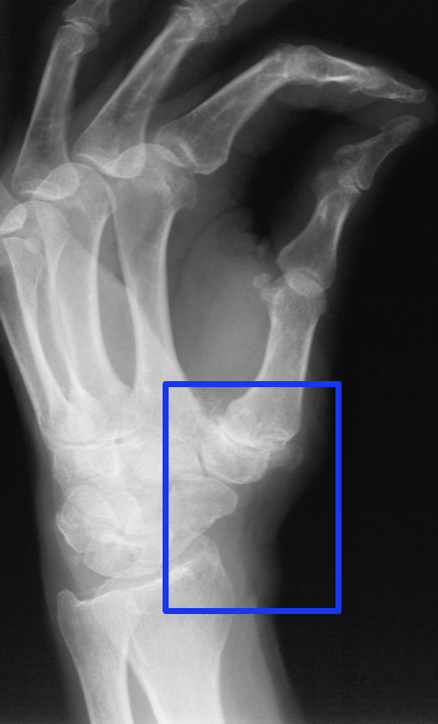
In osteoarthritis of the carpometacarpal joint, also known as rhizarthrosis, the cartilage on the joint surface of the trapezium bone and the base of the first metacarpal bone wears away, ultimately resulting in bone-on-bone contact. Sharp, bony outgrowths (osteophytes) sometimes form at the joint margins. These changes cause pain when gripping objects in this area, often radiating into the thumb and forearm. Holding heavy objects becomes nearly impossible, and even opening twist-off lids or turning a key becomes increasingly difficult.

SURGICAL TREATMENT
In the early stages of osteoarthritis, severing pain-conducting nerve fibers (joint denervation) or performing a realignment osteotomy can alleviate symptoms. However, this does not halt the natural progression of osteoarthritis. In some cases, though, the procedure delays the actual rhizarthrosis surgery by several years.
The most frequently chosen surgical treatment for advanced osteoarthritis is suspension arthroplasty. This involves removing the trapezium bone, which forms the proximal articular surface of the thumb's carpometacarpal joint. The goal of the procedure is to restore good, pain-free thumb movement. Therefore, a sufficiently long course of physiotherapy and occupational therapy is essential afterward. Other surgical treatment options include partial removal of the trapezium bone, joint fusion, and total joint replacement.
AFTERTREATMENT
After surgery, the thumb is immobilized in a plaster splint for 2-3 weeks. Then, physiotherapy exercises using the splint and occupational therapy can begin. These exercises should focus on abduction and flexion, as well as opposition. After 6 weeks, gradually increasing strength training for the thenar muscles is started.
Due to the removal of the carpal bone, strength is somewhat reduced after the operation compared to the healthy side. However, with proper training, a significant increase in strength is possible, allowing the hand to be used normally and painlessly in daily work.